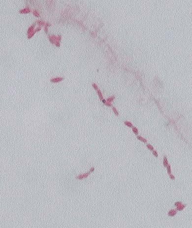
Simple stain of S. thermophilus
Growth on HYA agar:
After the first
incubation period, there were small colonies present on the agar.
We then restreaked isolated colonies onto HYA and TSA plates and
incubated again at 37 degrees Celsius. Every time, the colonies appeared
to be the same, and appeared to be the only type growing on the media.
The colonies were very small (between .5 and 1 millimeter in diameter),
and white, which is typical of S. thermophilus. However, L.
bulgaris is also expected to grow on this media, so the following tests
were performed to determine which organism was present. There was
a problem in differentiating the two because they have very similar testable
characteristics.
|
|
|
|
|
| Gram Stain | gram positive | gram positive | gram positive |
| Catalase | negative | negative | negative |
| Glucose Fermentation | positive | positive | positive |
| Mannitol Fermentation | negative | negative | negative |
| Simple Stain | inconclusive: appears as if both rod shaped and ovoid shaped bacteria were on slide. The small size of the bacteria made it hard to determine a shape. | spherical or ovoid shaped, in chains | rod shaped |
Photos:
Simple stain of S. thermophilus
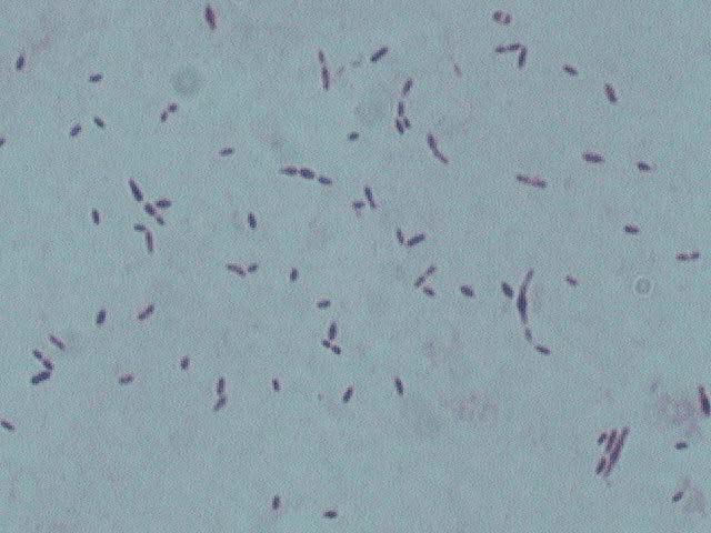
Gram stain of S. thermophilus-
purple color indicates gram positive organism
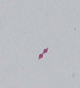
Above, left is light microscope image
of a simple stain of S. thermophilus completed in this experiment.
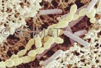
Above, right is an electron microscope picture of S. thermophilus
and L. bulgaricus. Note the lemon-like appearance of S.
thermophilus chains that would make it hard to be distinguished from
the L. bulgaricus rods. (Photo from: http://anka.livstek.lth.se:2080/microscopy/f-yogurt.htm)
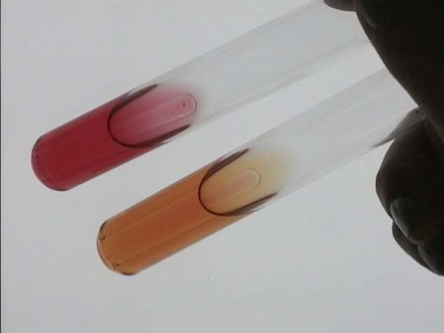
Sugar tubes innoculated with isolated
organism: top is mannitol (not fermented), bottom is glucose (fermented).