The Poly-Vagal Theory is based on several premises. Some are firmly grounded in neurophysiological and neuroanatomical data and others are more speculative. The first premise articulates the neural regulation of bradycardia and RSA. Based upon the initial premise, it is hypothesized that the neurogenic bradycardia associated with the orienting reflex are mediated by DMNX and that the suppression of heart rate variability (i.e., reduced amplitude of RSA) is mediated by NA.
Premise 1: Neurogenic bradycardia and RSA are mediated by different branches of the vagus and need not respond in concert.
Physiological support for the hypothesis that DMNX can contribute to neurogenic bradycardia, independent of NA, is provided by lesion studies. Machado and Brody (1988) have reported that chronic bilateral lesions of NA reduced but did not totally block baroreceptor reflex-mediated bradycardia in conscious rats. Thus, DMNX contains vagal neurons capable of producing bradycardia with a response latency associated with the baroreceptor reflex. This is supported by Jerrell, Gentile, McCabe, & Schneiderman (1986), who argued that differential Pavlovian conditioning of bradycardia in rabbits, following sinoaortic denervation, was mediated via DMNX pathways. The results pose the possibility that vagal pathways, originating in both the DMNX and NA, have the potential to influence heart rate.
Phylogenetic development of the Poly-Vagal System
Investigations of the phylogenetic development of the vagus provide support for the first premise. Since our interests are in mammals and specifically humans, this paper will focus on the evolution of vagal regulation of cardiac function from reptiles to mammals. There are two questions: 1) Do reptiles produce heart rate patterns during orienting similar to the neurogenic bradycardia observed in mammals? 2) Do reptiles produce a phenomenon similar to RSA?
The phylogeny of the vagus illustrates two phenomena: one, neuroanatomical, and the other, physiological. On a neuroanatomical level, differentiation of the visceral efferent column of the vagus into a dorsal motor nucleus (i.e., DMNX) and a ventrolateral motor nucleus (i.e., NA) is first seen in reptiles. In turtles (e.g., Chelone mydas and Domonia subtrijuga) there is still a connection between the two nuclei, but in lizards (e.g., Varanus salvator) and crocodiles (e.g., Caiman crocodilus) the separation between DMNX and NA is as complete as it is in mammals (see Barbas-Henry & Lohman, 1984).
Behavioral orienting in reptiles is characterized by a focusing of exteroceptors and a freezing of gross motor activity. Paralleling these behaviors, neurogenic bradycardia have been observed. Belkin (see Regal, 1978) reported that bradycardia is part of a fear response in iguanas. Additionally, McDonald (1974) reported brady-cardia in the hog-nosed snake during death feigning. Most researchers found these data incompatible with the prevalent emphasis on arousal and the use of heart rate as indicator of arousal. How could bradycardia reflect increased arousal within the context of a sympathetic nervous system oriented arousal theory? In contrast, RSA has not been observed in reptiles. Research investigating the spectral components of reptilian heart rate has failed to identify heart rate oscillations associated with ventilation (Gonzalez Gonzalez, & de Vera Porcell, 1988).
Phylogenetic development not only illustrates changes in the neuroanatomy of the vagus, but also parallel changes in behavior. One of these behavioral shifts is the addition of active or voluntary attention and complex emotions. In confronting the defensive world, mammals, like reptiles, have an initial reflexive response to novelty, the orienting reflex. However, mammals have additional behaviors in their repertoire. Following or independent of reflexive orienting, mammals may voluntarily respond with sustained attention to foster detailed information processing, or with facial expressions and vocalizations to foster communication. Thus, reptiles orient; mammals may first orient and then elect to attend or communicate.
The differences between the reptilian and mammalian cardiac systems provides insight into the phylogenetic differences in behaviors such as reptilian orienting and mammalian attention and emotion. The cardiac output and thus, energy production of mammals far exceeds that of reptiles. Mammals have metabolic demands four to five times that of reptiles. The metaphor of a machine or vehicle has been proposed by Else & Hulbert (1981) to compare the efficiency and function of the mammalian and reptilian metabolic systems. According to Else and Hulbert (1981), when idling, the average mammal requires four to five times more fuel than the average idling reptile, even when body weight and ambient temperature are controlled. Elaborating on this metaphor, reptiles represent vehicles with one-liter engines and mammals represent vehicles with four- or five-liter engine. Thus, as in the story about the race between the tortoise and the hare, reptiles locomote with a reliable but under-powered engine and mammals locomote with a supercharged engine that can function for only short periods without requiring refueling.
The energy production capacities of reptiles and mammals contribute to their respective lifestyles. There is a bias among reptiles toward passive feeding strategies. Reptiles tend to be sit-and-wait feeders, slow cruisers, and sluggish browsers. In contrast, mammals with four-chambered hearts, can actively hunt and graze and adapt to changing environments (Regal, 1978). To support their behavioral niche and to ensure their adaptive success, reptiles and mammals use different vagal strategies to promote their lifestyles. Being under-powered, reptiles do not maintain a vagal brake on the heart, which would further reduce energy production during unchallenged situations. For reptiles, during periods of either quiescence or apnea, usually associated with behavioral freezing or diving, vagal influences via DMNX are profound and heart rate is even slower. In contrast, vagal control of the heart is virtually removed during periods of breathing and other motor activities (Belkin, 1964; Jacob & McDonald, 1976).
The under-powered reptiles use vagal efferents from DMNX to the heart to deal with specific challenges: to orient and freeze in response to predator or prey, and to conserve oxygen while submerged for lengthy periods. In contrast to under-powered reptiles, supercharged mammals use vagal efferents from NA as a persistent brake to inhibit the metabolic potential of this high-powered system. The high NA-vagal tone keeps mammals from, literally, bouncing off walls. Thus, in contrast to that observed in reptiles, in mammals vagal tone is highest during unchallenged situations such as sleep, and vagal tone is actively withdrawn in response to external demands, including metabolically demanding states such as exercise, stress, attention, and information processing. For example, in humans, psychological states perceived as life threatening, such as panic and rage, are characterized by virtually no NA-vagal tone when indexed with the amplitude of RSA (George et al., 1989). Metaphorically, and consistent with the model, antisocial and pathological behavioral patterns associated with rage and hyper-reactivity without conscious self-regulation, have been labeled reptilian.
If terrestrial mammals adopted the reptilian strategy of reflexive increases in vagal activity to produce massive neurogenic bradycardia, the result would be catastrophic to the oxygen-hungry mammalian cortex and myocardium. This strategy would rapidly produce cardiac ischemia and cortical anoxia. The result of this sequence would be death. Although still dependent on oxygen, aquatic mammals use a diving reflex characterized by a regulated neurogenic bradycardia to reduce metabolic demands. To survive, aquatic mammals have complex mechanisms, not available to terrestrial mammals, to manage oxygen resources and shift priorities for oxygen while submerged for long periods.
It is possible that for mammals, during states of stress, when metabolic demands are great and vagal tone from NA is removed, that the cardiac pacemakers (S-A and A-V) may be prone to neurogenic bradycardia mediated by DMNX. The neurogenic bradycardia may be massive and lethal. This may be the case in fetal distress, when bradycardia are observed during hypoxic episodes, or as a factor in either sudden infant death syndrome (SIDS) or sudden death in adults. Consistent with this model, it has been demonstrated in the dog that progressive asphyxic hypoxia, not only elicits increased cardiac vagal activity, but the sensitivity of the sino-atrial node to vagal efferent influences is potentiated (Potter & McCloskey, 1986). Thus, during hypoxia large bradycardia may be maintained with limited or reduced vagal efferent activity.
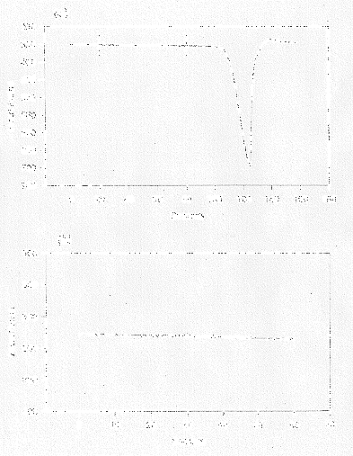
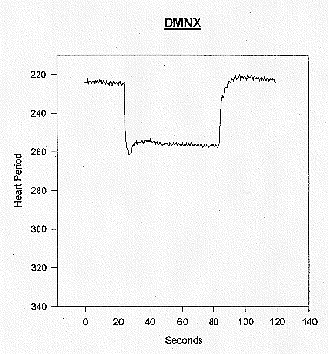
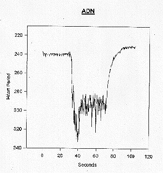
The Poly-Vagal Theory provides a potential explanation for the massive neurogenic bradycardia observed during fetal distress and in high risk neonates who have virtually no observable RSA. For example, as illustrated in Figure 2, when massive bradycardia are observed during fetal distress (a), there is a background of low beat-to-beat variability (b). Similarly, neonates with the lowest amplitude RSA are at greatest risk for apnea and bradycardia (Sostek, Glass, Molina, & Porges, 1984). Thus, the diminished vagal influences from NA, responsible for depressed RSA amplitude, seem to be associated with a vulnerability to large neurogenic bradycardia. Potter and McCloskey (1986) provide an explanation of how depressed CNS function associated with hypoxia might result in massive neurogenic bradycardia. They report a complex feedback system between duration of hypoxia, vagal efferent discharge, and potentiation of the vagal output on the heart. This system is able to maintain bradycardia, despite the massive decline in the vagal firing associated with hypoxia, by potentiating the influence of the vagal firing on the S-A node. Under these conditions, although the bradycardia are mediated through a branch of the vagus, the magnitude of the bradycardia is determined by a peripheral mechanism and no longer reflects a centrally mediated vagal tone. Although Potter and McCloskey did not monitor RSA, we must assume that RSA is low in their preparation because the animals were anesthetized prior to the surgical, electrical, and hypoxic manipulations, and because both hypoxia and anesthesia are associated with depressed beat-to-beat heart rate variability, including RSA (e.g., Donchin, et al., 1985; Nelson, 1976). Additional support for this bifurcation of vagal influences is demonstrated by electrical stimulation of the dorsal motor nucleus in the rabbit. As illustrated in Figure 3, electrical stimulation of DMNX results in a bradycardia without an increase in RSA. This is in contrast to the effect of stimulation of the aortic depressor nerve, which com-municates with both NA and DMNX. Note in Figure 4 that in a similar anesthetized rabbit, stimulation of the aortic depressor nerve results in an increase in RSA and a massive bradycardia (e.g., McCabe, Yongue, Porges, & Ackles, 1984).
The Poly-Vagal Theory argues that the vagal fibers from the DMNX and NA are distinguishable in structure and function. Specifically, it has been argued that the vagal efferent fibers from the NA are myelinated and contain a respiratory rhythm and the vagal efferent fibers from the DMNX are unmyelinated and do not express a respiratory rhythm. However, there are some inconsistencies in the proposed distinction. For example, as cited above, Jordan et al. (1982) reported that there are cardioinhibitory vagal neurons originating in the DMNX with efferent axons conducting in the B fiber range and therefore, myelinated. Moreover, Jordan et al. report that these had a respiratory rhythm. Although the Jordan et al. findings support the proposed dual source of vagal efferents, their findings confound the proposed functional distinction.
There are several potential explanations for the inconsistency identified by Jordan et al. (1982). First, may be method. The Jordan et al. study used standard neurophysiological stimulation and recording techniques to identify cell bodies. According to Schwaber (1986) many vagal fibers previously assumed to originate in DMNX have been identified with newer methods, such as retrogradely labeled HRP, to be located in NA. Schwaber (1986) also states that since axons from the NA pass very near the lateral border of DMNX, it is difficult to stimulate or lesion DMNX without NA involvement and confounding electrical stimulation studies. Thus, additional research with more accurate labeling techniques may demonstrate that all neurons in the B fiber range originate in NA. Our rabbit data reported in Figures 3 and 4 provide additional support for the possibility of mislabeling. According to Jordan et al. (1982) all neurons excited by aortic depressor nerve stimulation produced a respiratory rhythm in their ongoing discharge. Similarly, as illustrated in Figure 4, stimulation of the aortic depressor nerve resulted in both bradycardia and increased RSA. However, stimulation of the DMNX produced only an attenuated bradycardia. These findings suggest that vagal fibers discharging following stimulation of DMNX did not have a respiratory rhythm. Moreover, the bradycardia was immediate and similar in latency to that observed following aortic depressor nerve stimulation. However, the magnitude of bradycardia was about 50% of the magnitude elicited via aortic nerve stimulation that is assumed to recruit vagal fibers from both NA and DMNX. These findings are similar to those reported by Machado and Brody (1988). A second possibility is that there are species differences in the organization and function of the DMNX. For example, to facilitate freezing behavior, the rabbit may have evolved unique myelinated vagal pathways from the DMNX that were independent of respiratory function. According to this explanation, the DMNX would have B fibers, but they would express a respiratory rhythm. Alternatively, some mammalian species may have neurons in or near the DMNX that are part of a common cardiopulmonary oscillator (Spyer & Richter, 1990).
Future research will determine whether the proposed functional and structural distinctions between DMNX and NA efferents articulated in the Poly-Vagal Theory are accurate. An additional concern relates to generalizing across mammalian species. Most of the neurophysiological and neuroanatomical research on the mammalian vagus has been conducted with rat, rabbit, cat and dog. Studies investigating vagal regulation with humans have been limited to pharmacological blockade studies with measures of peripheral physiology. There are few neuroanatomical studies of human brain stem, however, these studies are often conducted on patients who have died of disease or trauma. Thus, one may question the generalizability of a poly-vagal model developed from investigations of rodent brain stem to the human. However, existing data illustrates phenomena such as clinical bradycardia in the absence of RSA in the human fetus (see Figure 2), shifts in RSA independent of heart rate change during inhalant anesthesia (e.g., Donchin et al., 1985), and short latency responses from both systems (see Figures 3 and 4) that argue for a poly-vagal system.
Vagal strategies in mammals and reptiles
Reptilian and mammalian vagal systems have contradictory strategies. Reptiles are characterized by low ambient vagal tone and transient increases in vagal tone in response to environmental challenges. In contrast, mammals are characterized by high ambient vagal tone and transient decreases in vagal tone in response to environmental challenges.
Table 3
Vagal Strategies
To adapt to the hostile world, the reptiles' behavioral repertoire is survival driven. Most behaviors are associated with foraging, stalking, and feeding. Only limited time and energy are dedicated to social interactions such as parenting and reproduction. In the reptiles' defensive world, neurogenic bradycardia are adaptive and do not compromise physiological status. Reptiles have smaller metabolically active body organs, have different metabolic mechanisms, are less oxygen dependent than mammals, and can go for long periods without oxygen. In contrast, the adaptive strategy of reptiles is lethal for mammals. In the defensive world of mammals, it is necessary to increase metabolic output to foster fight or flight behaviors. Therefore, reflexive neurogenic bradycardia to novelty for a prolonged period would reduce oxygen resources and metabolic output and compromise the fight or flight potential of mammals. The consequences of reduced oxygen resources also would depress central nervous system function, reduce behavioral complexity and competent execution of complex behaviors, induce unconsciousness, damage vital organs and finally, if persistent, result in death. Thus, the cardiac component of the orienting reflex must be of short duration and replaced by a physiological response that does not compromise the oxygen-needy nervous system of mammals. The withdrawal of vagal tone via NA serves this purpose.
Phylogenetic origins of vagal response patterns
The neurogenic bradycardia controlled by DMNX and observed in reptiles and mammals during orienting may have evolved from the gustatory response system of primitive vertebrates. Gustation is the primary method for identifying prey (including other appropriate food sources) and predators in aquatic environments. For example, in fish, an undifferentiated vagal lobe controls gustatory, digestive, and alimentary processes (Finger & Dunwiddie, 1992). A reflexive increase in vagal tone would affect several organs: 1) the heart, where it would reduce metabolism and enable the animal to freeze momentarily, 2) the organs containing gustatory receptors, where it would orient towards the source of stimulation and regulate threshold to detect novelty, and 3) the digestive and alimentary systems, where it would stimulate gastric secretion and motility.
With phylogenetic development, the viscerotropic organization of the vagal system has become more complex, and incorporates pathways from other cranial nerves including trigeminal, facial, accessory and glossopharyngeal. Thus, more specialized functions such as head rotation to orient sensory receptors toward the source of stimulation, mastication to ingest food, and salivation to initiate gustatory and digestive processes are integrated into the vagal system.
The motor component of the vagus shares evolutionary origins with the four cranial nerves mentioned above (trigeminal, facial, accessory and glossopharyngeal). The vagus not only innervates smooth and cardiac muscle, but similar to the other four cranial nerves, it contains motor pathways that innervate somatic muscles. Vagal pathways that innervate somatic muscle often are not included in the neurophysiology of the autonomic nervous system. These fibers are labeled special visceral efferents to distinguish them from the motor pathways innervating smooth and cardiac muscle that are labeled general visceral efferents. The critical difference between the two types of motor pathways is that somatic muscle regulation may be conscious and voluntary, while smooth muscle regulation is reflexive and unconscious. Since the special visceral efferents innervate voluntary muscles, usually they are excluded from the autonomic nervous system. Traditionally, only the general visceral efferents from both sympathetic and parasympathetic branches are used to define the autonomic nervous system.
The somatic muscles innervated by the five cranial nerves arise from the branchial arches, embryologically known as the primitive gill arches, (Warwick & Williams, 1975). These muscles are critical to several mammalian behaviors. For example, the somatic muscles innervated by the trigeminal, arising from the first branchial arch, are involved in mastication, retraction of the lower jaw, and closing the mouth, The special visceral efferents from the facial nerve, arising from the second branchial arch, innervate the muscles of the face, scalp, and neck to enable facial expressions. The facial nerve also innervates muscles in the floor of the mouth. Although the trigeminal and facial nerves originate from branchial arches and have communications with the other three cranial nerves originating from the branchial arches, the source nuclei of the special visceral efferents for the glossopharyngeal, vagus, and accessory nerve originate in the same medullary nucleus, the nucleus ambiguus (NA). Thus, the efferent fibers travel through three different cranial nerves, but they originate in the same source nucleus.
As a function of phylogenetic development, the source nuclei for the special visceral efferent pathways in the glossopharyngeal, vagus, and accessory nerves migrate to form NA. In mammals, NA controls the complex coordination of pharynx, soft palate, larynx, and esophagus. Of special note to psychophysiological processes, the third gill arch also gives rise to the carotid body, containing peripheral chemosensitive receptors sensitive to oxygen and carbon dioxide levels, (Warwick & Williams, 1975). In addition, the accessory nerve provides fibers originating in the cervical spinal cord that innervate the positioning of the neck. The critical carotid arteries, internal jugular veins, and vagus nerves run deep in these muscles (Warwick & Williams, 1975). Thus, this complex also has the ability to orient visceral receptors via somatic muscles, to coordinate structures related to ingestion and expulsion, and to regulate facial expression and emotion. These motor nuclei receive input from cortex to coordinate these complex behaviors with cardiopulmonary function. Thus, phylo- genetically, even when the gill arches evolve into the branchiomeric muscles common to all mammals, oxy-genation of blood through a coordination of breathing and heart rate during interactions with the environment remains a primary functional objective.
The processes associated with NA control of supradiaphragmatic organs appear to be uniquely mammalian. For example, this subsystem of the vagus coordinates the complex sequence of sucking, swallowing, and breathing that allows mammals to actively and voluntarily feed and breathe. Moreover, NA provides the primary chronotropic control of the heart and controls the intonation of vocalizations. Thus, NA efferent projections are involved with processes associated not only with feeding and breathing, but with processes associated with movement, emotion, and communication. These behaviors contribute to the unique social and survival behaviors observed in mammals. The NA-vagus provides the vagal brake that mammals remove instantaneously to increase metabolic output to foster fight or flight behaviors. The NA-vagus provides the motor pathways to shift the intonation of vocalizations (e.g., cry patterns) to express emotion and to communicate internal states in a social context.
The behavioral derivatives of the two branches of the vagus, suggest a typology in which one branch of the vagus deals with unconscious reflexive vegetative functions and the other branch of the vagus is involved in more conscious, voluntary, flexible and often social activities. There is neuroanatomical support for this typology. DMNX contains only general visceral efferents that innervate smooth and cardiac muscle fibers and regulate glandular secretion. In contrast, NA contains special visceral efferents that innervate the somatic musculature of the soft plate, larynx, pharynx, and esophagus.
Somatomotor and Visceromotor: Coupled systems
In mammals, we observe two evolutionary strategies that link autonomic function with somatic muscle activity. First, there is an anatomical linkage between the segmentation of the spinal nerves and the sympathetic chain. This linkage is reflected in the motor-related increases in sympathetic tone that have dogged psychophysiologists by confounding motor and autonomic responses. The evolution of the segmented sympathetic nervous system parallels the evolution of voluntary motor activities. The sympathetic nervous system regulates vasomotor tone to direct blood flow, and thus, oxygen, to the specific muscles being challenged. Additionally, there are sudomotor links to hydrate and protect the skin from tearing. This link between sympathetic activity and movement has been the cornerstone of arousal theory and hypotheses linking autonomic function to temperament and psychopathologies. It was not many years ago that Obrist challenged the Lacey notion that autonomic state was independent of motor activity (i.e., metabolic demands). There is no doubt that the effects of motor activity are profound on the autonomic nervous system. Yet, this profound effect does not mitigate the importance of other relationships that may be sensitive to specific psychological processes, independent of movement.
Second, there is an anatomical linkage between the somatic muscles that arise from the cranial nuclei and parasympathetic function. We can observe this clearly in the viscerotropic organization of NA. NA provides the source nuclei for somatic muscle fibers that innervate larynx, pharynx, trachea, and esophagus. Moreover, ventral to these source nuclei, in an area of the nucleus ambiguus known as NAex, are general visceral efferents that control the resistance of the bronchi (Haselton, Solomon, Motekaitis & Kaufman, 1992) and heart rate (Bieger & Hopkins, 1987). The ventral portion also projects to other visceral organs (e.g., Brown, 1990).
Based upon neuroanatomical studies, it has been demonstrated that visceromotor functions regulated by the ventral part of NA provide the parasympathetic support for the somatomotor projections from NA, trigeminal and facial nerves. Neuroanatomical studies suggest that, unlike DMNX, which receives primary sensory input via NTS, NA has the trigeminal nerve as an important source of sensory input. Moreover, the rostral region of NA communicates with the facial nucleus. This coupling of NA with facial and trigeminal nuclei provides additional evidence of the coordination of the visceromotor regulation via NA with somatomotor functions such as swallowing (Brown, 1974), sucking (Humphrey, 1970), and, perhaps, facial expressions. Thus, the organization of the mammalian brain stem has evolved to have a ventral vagal complex consisting of NA and the nuclei of the trigeminal and facial nerve that co-exists with the dorsal vagal complex consisting of The DMNX and NTS that regulates vegetative processes and is observed in the reptile.
To foster motor movement, visceromotor (i.e., autonomic) processes are associated with somatomotor activities. In the periphery this is done primarily by the sympathetic chain and in special cases, such as those related to reproduction and elimination, the sacral branch of the parasympathetic nervous system contributes. However, in the rostral part of mammalian anatomy (i.e., the head) the somatic muscles that regulate facial expression, mastication, vocalization, swallowing, and sucking are matched with general visceral efferents, projecting from the ventral portion of NA, that exert potent influences on the heart and the bronchi. These motor fibers effectively slow heart rate and increase respiratory resistance to conserve oxygen exchange. Neuroanatomical studies performed on human embryos and fetuses suggest that these visceromotor neurons may have migrated from DMNX (Brown, 1990).
As observed through both embryological research and phylogenetic comparisons, in mammals, the primitive gill arches evolve into muscles and nerves controlling the face, bones of the mouth, jaw, pharynx, larynx, softplate, esophagus, and trachea. The nerves innervating these muscles uniquely arise, not from the anterior horns of the spinal cord, but from the source nuclei of five cranial nerves referred to above (trigeminal, facial, glosso-pharyngeal, vagus and accessory). Because of their uniqueness, these motor systems are known as special visceral efferents. And, because of their voluntary aspects, these pathways have been excluded from traditional concepts of the autonomic nervous system. Facial expressions, sucking, swallowing, and vocalizations, characteristic of mammals, reflect the unique mammalian adaptation of special visceral efferent control of the visceral muscles evolving from the branchial arches.
However, similar to the synergistic relationship between the sympathetic nervous system and skeletal muscles of the extremities, there is a synergistic relationship between the traditional general visceral efferents of the vagus and the somatic muscles controlled by these cranial nerves. Thus, increased outflow of these somatic muscles produce specific visceral shifts. For example, chewing will produce salivation in the absence of food. Additionally, head rotation, via accessory special visceral efferents, will impact on cardiovascular action via the vagus.
Phylogenetic development of the central nervous system has progressed in mammals to produce a brain with a large neocortex (e.g., MacLean, 1990). The neocortex is very vulnerable to shifts in oxygen. Evolutionary pressures have resulted in autonomic strategies that optimize the availability of oxygen to the cortex. However, these uniquely mammalian strategies coexist with the ancestral reptilian strategies. Thus, premise 2 is stated, consistent with MacLean's view that the advanced mammalian brain contains its phylogenetic heritage.
Premise 2: Neurogenic bradycardia associated with orienting are a phylogenetic vestigial relic of the reptilian brain and are mediated via DMNX.
Although phylogenetic development has modified several brain structures, the evolved brain of advanced mammals maintains several structures and systems that are virtually identical to those observed in primitive reptiles. These primitive structures have extensive interconnections and functional dependencies, although each is capable of specific independent functions. Thus, in mammals, DMNX still maintains its reptilian functions of facilitating digestion and slowing heart rate. Mammals utilize an additional brain stem structure, the NA, to supply general visceral vagal efferents that provide the prominent control of the heart and the bronchi. The cells of origin of these fibers efficiently communicate with limbic, and other higher, centers and allow for the conscious and voluntary selection of novelty. In contrast, DMNX is more directly regulated by hypothalamic communication, often triggered by survival-oriented stimuli (Hopkins, 1987; Leslie, Reynolds, & Lawes, 1992). Thus, as stated in Premise 3, the regulation of vagal efferents by NA mechanisms contributes to the mammalian ability to detect novelty, actively engage with the environment, and socially communicate.
Premise 3: Withdrawal of cardiac vagal tone via NA mechanisms is a mammalian adaptation to select novelty in the environment, while coping with the need to maintain metabolic output and continuous social communication.
To summarize the reptilian-mammalian evolutionary evidence, phylogenetic development of the neural regulation of the heart provides insights into an apparent contradiction or paradox in vagal control of the heart. In most reptiles the neuroanatomy demonstrates: 1) a lack of anatomically distinguishable boundaries between DMNX and NA, and 2) cardiac vagal efferent pathways originating only in DMNX. In mammals the neuroanatomy demonstrates: 1) a distinct separation of DMNX and NA, 2) cardiac vagal efferent pathways originating primarily (but not exclusively) in NA, 3) direct neural connections between the central nucleus of the amygdala and NA, and 4) a clustering of medullary neurons in NA capable of regulating the somatic muscles related to vocalizations, facial expression, and to coordinate breathing with sucking and swallowing.
Smart and vegetative vagi.
The Poly-Vagal Theory proposes that the evolutionary shift resulting in both a NA that is distinct from the DMNX, and the evolutionary development of special visceral efferents changed the role of the vagus. The general visceral efferent pathways from DMNX vagus are part of a passive reflexive motor system associated with vegetative function and thus, a vegetative vagus. The special visceral efferent pathways from NA create an active voluntary motor system associated with the conscious functions of attention, motion, emotion, and communication, and thus, a smart vagus.
The Poly-Vagal Theory requires a reconcep-tualization of the vagal system and the construct of vagal tone. The Theory focuses on the cytoarchitecture of the medullary source nuclei of the cranial nerves. The Theory takes an evolutionary approach and investigates, via embryology and phylogenetic comparisons, the common origins of the special visceral efferents and focuses on the shared medullary structures for the cell bodies of these fibers. The Theory acknowledges that the vagal system is complex, and should be organized, not in terms of bundles of fibers leaving the medulla, but rather in terms of the common source nuclei of several of these pathways. Functionally, the common source nuclei both provide a center to coordinate and regulate the complex interactions among various end organs and are related to optimizing cardiopulmonary function.
Mammals, with their oxygen-hungry metabolic systems, require a special medullary center to coordinate cardiopulmonary functions with behaviors of ingestion (e.g., mastication, salivation, sucking, swallowing), oral or esophageal expulsion (vomiting), vocalizations (e.g., cries, speech), emotions (e.g., facial expressions), and attention (e.g., rotation of the head). The NA plays this role and serves as the cells of origin of the smart vagus. The potent link between NA and cardiopulmonary function observed in mammals is not observed in reptiles. In reptiles, which do not have nerves to regulate facial expression, NA does not play a major role in viscero-motor regulation.
Medullary contributions to a common cardiopulmonary oscillator
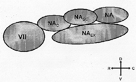
The NA is a continuum of interconnected sub-divisions beginning rostrally at the level of the facial nucleus and extending caudally to the spinal medullary junction. As illustrated in Figure 5, the rat NA has several subdivisions. The subdivisions are labeled: the compact (NAc), semicompact (NAsc), loose (NAl) and external (NAex) formations (Bieger & Hopkins, 1987). The dorsal division consists of NAc, NAsc, and NAl . The dorsal division is the source of special visceral efferents innervating the softpalate, pharynx, larynx, and esophagus. The ventral division consists of the NAex and is the source of general visceral efferents innervating the thoracic viscera, primarily the bronchi and the sino-atrial node. Vagal fibers originating in NAex and terminating in both the bronchi (Haselton et al, 1992) and the sino-atrial node (Spyer & Jordan, 1987) have a respiratory rhythm, thus suggesting that RSA may reflect a common respiratory rhythm originating in or, at least, incorporating NA.
After investigating the neuroanatomical centers associated with laryngeal, pulmonary, and cardiac function, Richter and Spyer (1990) arrived at a convergent conclusion that NA was a contributor to a common respiratory rhythm. They also speculated that mammals, with their great needs for oxygen, have a medullary center to regulate cardiopulmonary processes. They proposed that a common cardiorespiratory oscillator evolved to foster coordination among cardiac and respiratory processes. In their model the respiratory rhythm is dependent on the interaction between two groups of neurons, one in NTS and the other in NA. Accordingly, the "common" oscillator producing respiratory frequencies is a manifestation of a neural network composed of interneurons between areas containing the motoneurons regulating respiratory, laryngeal and cardiac function. Note that the cardiorespiratory oscillator does not involve DMNX. To support their hypotheses they report cross-correlational studies of single units. Thus, NA is part of the cardiorespiratory oscillator network, and the period of the oscillations in heart rate, (the period of RSA) provides a valid index of the output frequency of the cardiopulmonary oscillator.
Other researchers emphasize the importance of additional brain structures as contributors to the regulation, if not the generation, of a cardiopulmonary rhythm. For example, Harper and associates have demonstrated that "respiratory rhythms" can be observed in several nuclei in the brain stem, midbrain and forebrain. Harper's group, employing crosscorrelation techniques, has reported units firing on a breath-by-breath basis in periaqueductal gray (Ni, Zhang & Harper, 1990), central nucleus of the amygdala (Frysinger, Zhang, & Harper, 1988), hippocampus (Frysinger & Harper, 1989), and the anterior cingulate (Frysinger & Harper, 1986). In addition, they have reported that stimulation of amygdala can influence the respiratory cycle (Harper, Frysinger, Trelease, & Marks, 1984).
The link between bronchi and heart rate oscillations (e.g., RSA) mediated via NA may have a functional influence on the oxygenation of blood. As stated above, the primary objective of the phylogenetic derivatives of the primitive gill arches is to maintain oxygenation. Thus, one might speculate that oscillations in vagal tone to the bronchi and the heart might influence oxygenation. Perhaps, coherent rhythmic shifts between bronchial tone and heart rate, with a fixed phase lag, maximize oxygen diffusion. To answer this question, research would need to confirm a relationship between oxygen saturation and RSA, independent of the average heart rate and respiration rate. Currently, only anecdotal data exist that demonstrate that clinical conditions in which oxygen saturation is low, tend to be conditions in which RSA also is depressed. Support for this hypothesis is obtained from research demonstrating that vagotomy disrupts the oxygen consumption-oxygen delivery relationship (Schertel, Brourman, Klilng, Schmall, Tobias & Myerowitz, 1994).
Measurement of NA status: Quantification of RSA
For psychophysiologists, our interest is primarily in the behaviors and psychological processes associated with special visceral efferents. Most research has been directed toward processes that require the ability to monitor and mediate complex behaviors, such as attention, motion, emotion, and communication; these processes are neurophysiologically linked to the special visceral efferents of NA, facial, and trigeminal nerves. Yet, many of us measure only general visceral efferents from both the parasympathetic and sympathetic branches, although we are interested in the special visceral efferents that regulate vocalizations and facial expression. We are not at a total loss, because there is interneuronal communication between the dorsal and ventral segments of NA. Thus, by the nature of the NA having the general visceral efferents regulating heart and bronchi, it is possible to monitor continuously the vagal output or tonus of the smart vagus. This leads us to the fourth premise of the Poly-Vagal Theory.
Premise 4: The ability of NA to regulate both special visceral efferents and general visceral efferents may be monitored by the amplitude of RSA.
The vagal fibers originating in the NAex have a characteristic respiratory frequency that reflects a waxing and waning of influence. For example, the vagal fibers from NA that have an inhibitory action on the sino-atrial node also wax and wane in inhibitory influence at the respiratory rhythm and produce RSA. Thus, it is possible to monitor continuously the general status of NA by evaluating RSA. Similarly, NA fibers to the bronchi that elevate lung resistance also wax and wane in their inhibitory influence (Haselton et al., 1992).
RSA is a measure of the general visceral efferents of the NA, and thus is an index of the smart vagus. RSA is not a global measure of vagal tone or even a measure of "total" vagal control of the heart as previously proposed (Fouad, Tarazi, Ferrario, Fighaly, & Alicandro, 1984; Katona & Jih, 1975; Porges, 1992). There are other vagal and non-vagal influences on the heart, which contribute to both heart rate level and rhythm. For example, there are DMNX projections, as well as monosynaptic cholinergic pathways within the heart, sympathetic pathways, and intrinsic factors. However, the primary, if not sole, source of respiratory rhythms on the S-A node is due to projections from NA.
To evaluate NA regulation of the sino-atrial node, the parameters of RSA must be accurately extracted. We have approached this problem by evaluating the period and amplitude of RSA, independent of slower oscillations and trends, via a moving polynomial approach (Porges & Bohrer, 1990). In our research we have obtained correlations between respiration rate and period of RSA approaching 1.0. These findings support the notion of a common cardiorespiratory oscillator as described by Richter and Spyer (1990). RSA, with its amplitude representing visceromotor tone and its period representing the common cardiorespiratory drive frequency, is the functional consequence of the output of vagal fibers originating in NA and terminating on the sino-atrial node.
Quantification of RSA requires only an accurate determination of the amplitude and period of these oscillations. Additional experimental constraints to regulate breathing rates might confound the visceral-medullary feedback system which determines central respiratory rhythms. For example, since paced breathing requires an awareness of breathing parameters, cortical influences on brain stem structures might modulate the gain of the feedback and influence the amplitude of RSA. Also, paced breathing may shift respiratory parameters, such as rate, amplitude, inspiration-expiration ratio, inter-breath pause, and resistance, from brain stem setpoints. Data have been reported illustrating that paced breathing may influence RSA (Sargunaraj, Lehrer, Carr, Hochron, & Porges, 1994).
Various manipulations or conditions that depress special visceral efferents, such as inhalant anesthesia, have profound influences on RSA (Donchin et al., 1985). Recovery of function of special visceral efferents is paralleled by a recovery of RSA. In neurology, diagnosis is often based on the evaluation of the special visceral efferents. In our research, we noted that RSA amplitude before neurosurgery was an effective diagnostic of neurological recovery following neurosurgery (Donchin, Constantini, Szold, Byrne, & Porges, 1992). Additional neurological data demonstrate consistent depression of RSA in individuals who are diagnosed as brain dead (Mera, Wityk, & Porges, 1995).
High risk preterm neonates have problems coordinating breathing, sucking, and swallowing (i.e., processes regulated by NA). These infants have low levels of RSA (Porges, 1992). Many of these infants have severe bradycardia. The bradycardia are often paralleled by apnea, and a drop in available oxygen and may be assumed to reflect neurogenic vagal regulation via DMNX. Recall, that this response, to deal with decreased resources, is adaptive for reptiles, but potentially lethal for the human. This also is observed during fetal distress, when there is severe hypoxia associated with a loss of RSA and a pronounced neurogenic bradycardia (see above).
Vagal Competition and Autonomic Dysfunction
The concept of competition between sympathetic and parasympathetic inputs is well known. For example, Levy (Vanhoutte & Levy, 1979; Levy,1984) has clearly documented the ability of vagal efferents to inhibit sympathetic influences. Similarly, Berntson and associates have modelled the interactions between sympathetic and parasympathetic efferents to the heart (Berntson, Cacioppo, & Quigley, 1991). However, there may be a different type of competition, in which the two vagal branches are conveying contradictory information to the target organs. Since both vagal pathways are capable of regulating heart rate, there may be competition on the sino-atrial node. Due to the rate of acetylcholine degradation on the nodal tissue (Dexter, Levy, & Rudy, 1989) the continuous stimulation of the sino-atrial node by NA pathways may functionally protect the heart from massive neurogenic bradycardia mediated by DMNX. Thus, the observations of massive pathophysiological bradycardia in hypoxic fetuses and neonates, who have very low amplitude RSA, may reflect the loss of NA protection on the SA node. Similarly, sudden death following exercise might reflect a similar process associated with the depression of NA input to foster metabolic activity, and a surge of DMNX input in response to decreased oxygen resources.
The vagal competition hypothesis may be generalized and tested to explain other autonomic diseases such as asthma. The vagal competition hypothesis proposes that all target organs with smooth and cardiac muscle have dual innervation from both the DMNX and NA. Currently, this has been documented in animal preparations for heart, lungs, esophagus, and abdominal viscera including pancreas, liver, and stomach (Brown, 1990). However, as with the heart, the two vagal inputs may innervate in a contradictory manner. Just as a DMNX surge coupled with low RSA can result in sudden death, as in the examples above, bronchial asthma may be produced by a similar mechanism. In the case of asthma, NA efferent control of bronchi results in the bronchi exhibiting a rhythmic waxing and waning with breathing. This continuous stimulation of the bronchi by NA pathways may functionally protect the bronchi from pathophysiological DMNX influences. It is possible that without NA influences, the bronchi become vulnerable to vagal surges from DMNX. This would be an adaptive response for a primitive brain stem attempting to conserve oxygen, but it is lethal for the oxygen- hungry mammal. The asthma attack, similar to lethal neurogenic bradycardia, may be a product of a primitive vago-vagal reflex. In this type of reflex, not only do the motor fibers originate, but the afferent fibers terminate in DMNX. There is an anatomical basis for a monosynaptic vago-vagal reflex. There are reports that dendritic processes from DMNX neurons extend into the boundaries of NTS. Thus, vagal afferent fibers may com-municate directly with DMNX neurons (Neuheuber & Sandoz, 1986). Since afferents terminate in DMNX, the name "motor nucleus" is not accurate and a preferred designation, "dorsal nucleus of the vagus nerve" has been suggested (Nara, Goto, & Hamano, 1991). In most vagal reflexes involving the bronchi, the afferents terminate in NTS and influence NA to provide a fail safe feedback system.
Based upon the Poly-Vagal Theory, the assumption of vagal competition promotes the following testable hypotheses:
Nucleus ambiguus (Vagal) Protection Hypothesis: Vagal projections originating in NA and terminating in visceral organs provide tonic influences that promote health, growth and restoration.
Nucleus ambiguus (Vagal) Withdrawal hypothesis: Removal of the NA-vagal brake for short periods of time promotes metabolic output to foster locomotion. Removal of these influences for long periods places the organ at risk.
Emotion
The Poly-Vagal Theory provides a set of predictions regarding the relation between autonomic responses and emotion. Darwin carefully described facial expressions as the primary defining characteristics of emotion. The special visceral efferents, associated with the facial nerve, control movements of facial expression. Reptiles do not have facial muscles and cannot modulate facial expression. The facial nerve in mammals not only regulates facial muscles but interacts with NA and the vagal system. Thus, it is logical that emotional expression, which requires somatic muscles controlled by special visceral efferents, is linked to the visceromotor regulation of cardiopulmonary function via NA vagal efferents. Additionally, special visceral efferents originating in NA regulate the larynx and control intonation. Thus, the following premise is stated.
Premise 5: Emotion, defined by shifts in the regulation of facial expressions and vocalizations, will produce changes in RSA and bronchomotor tone mediated by NA.
As a construct, emotion is heterogeneous. Therefore, correlations between specific emotions and physiological states may be a function of the type of emotion. Even Darwin (1872) distinguished between primary or neurally based emotions and social or culturally based emotions. Darwin (1872) suggested that certain emotions have as their substrate an innate neural basis and, because these emotions are neurally based, they are universally expressed and understood across cultures. These primary emotions include anger, fear, panic, sadness, surprise, interest, happiness (ecstasy), and disgust (Ross, Homan, & Buck, 1994). Since the prevalent hypotheses suggest a strong physiological basis for primary emotions, we will focus here on relating primary emotions to the Poly-Vagal Theory.
There are two important aspects linking the Poly-Vagal Theory to the study of emotion: first, there is a parallel between cortical asymmetry and autonomic asymmetry; second, the branchial arches have evolved into the structures that mammals use to express emotion (i.e., facial muscles, larynx).
The literature documents the relationship between right brain function and primary emotions (Heilman, Bowers, & Valenstein, 1985). The medullary source nuclei and efferent pathways of the vagus also are lateralized with a right bias. The right NA via the right cardiac vagus provides the primary chronotropic output to the heart. The special visceral efferents, which provide the behaviors that are used to define emotion (facial expression, vocalization) also have a right bias and are linked neuroanatomically to the general visceral efferents originating in NA that regulate the bronchi and heart; organs that are assumed to be sensitive to emotion and stress. It is difficult to predict the influence of this right bias on actual facial expressions. Since the face is controlled by upper motor neurons that are crossed and lower motor neurons that are uncrossed (Rinn, 1984), facial expression may not be systematically lateralized. In fact, research on facial asymmetry and emotion has not been consistent. There have been reports of facial expressions not being lateralized, being lateralized on the left, and others being lateralized on the right (e.g., Hager & Ekman, 1985).
The functional dominance of the right side of the brain in regulating autonomic function and emotion may have implications for the specialization of motor and language dominance on the left side of the brain. The right sided responsibilities of regulating homeostasis and modulating physiological state in response to both internal (i.e., visceral) and external (i.e., environmental) feedback, may contribute to the development of motor and language functions on the left side of the brain.
A partitioning of central control of voluntary processes, independent of emotional-homeostatic processes would enable the individual to express complex voluntary levels of communication and movement, via the left side of the brain, and more intense emotional-homeostatic processes, via the right side of the brain. If these processes are lateralized, they might have a degree of autonomous regulation. This would enable simultaneous activation of global functions associated with emotional-homeostatic processes and language-voluntary movement processes.
Given the strong theoretical relationships between lateralized autonomic and hemispheric function and between the neurons that control RSA and the neurons that control facial expression and vocal intonation (see Figure 5), research should be directed at evaluating the relationship between RSA and the primary emotions. Recall that the source nucleus of the facial nerve is the border of NA and afferents from the trigeminal nerve provide a primary sensory input to NA. Thus, the Ventral Vagal Complex consisting of NA and the nuclei of the trigeminal and facial nerves is clearly related to the expression and experience of emotion.
Based upon the Poly-Vagal Theory, one would expect shifts in affective state to parallel RSA. For example, the elicitation of a negative primary emotion would result in a systematic withdrawal of vagal tone via NA to promote fight and flight like behaviors. In contrast, a shift to a more pleasant affective state would be associated with an increase in RSA. A study by Bazhenova (1995), emphasizing the dynamics of RSA change during the shifting affective states, supports this speculation. Bazhenova manipulated the affective state of infants and demonstrated that when an infant shifted to a more negative affective state, RSA decreased. Moreover, when the infant shifted to a more positive affective state, RSA increased above the affectively neutral baselevel.
The Poly-Vagal Theory does not neglect the important role of DMNX in the emotional experience. For example, DMNX is critical in the regulation of digestive polypeptides and gastric motility (Uvnas-Moberg, 1989), dimensions of physiological activity that parallel emotive experiences and stress. Consistent with the Poly-Vagal Theory, which emphasizes the importance of NA and VVC in overt emotional expressiveness and regulation, the Theory would acknowledge the importance of less conscious survival oriented processes that are mediated via Dorsal Vagal Complex consisting of NTS and DMNX. A complementary theory has been proposed by Uvnas-Moberg (Uvnas-Moberg, 1987, 1994). The Uvnas-Moberg theory emphasizes the role of DMNX in the regulation of gastrointestinal hormones and during emotional states including stress, hunger and satiety.