Lab 6 Precipitation
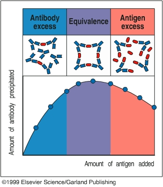
The precipitation reaction is a highly specific serological reaction involving the binding of antigen by antibody. Each antibody has two antigen binding sites, and each antigen could have multiple antigenic epitopes. When soluble antigens are bound by antibody to form a cross-linked lattice structure, the reaction is called precipitation. The precipitation reaction is inhibited when either antigen or antibody is present in great excess. When antibody is in great excess, separate antibody molecules may bind all available antigenic epitopes, preventing cross-linking. When antigen is in great excess both binding sites of antibody molecules will be bound with separate antigen molecules, again inhibiting cross-linking. The range of optimal concentration, at which the greatest amount of precipitation is generated, is called the equivalent zone. Based upon this principle, the presence of an antigen in a mixture and the relative concentration of an antigen can be detected using a specific antibody.
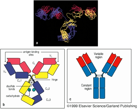
Antibody structure
Equivalent zone
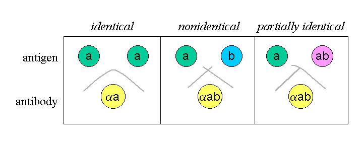
In this lab exercise, you will learn how to do Ouchterlony test. The Ouchterlony test is a double diffusion technique developed by Organ Ouchterlony more than 40 years ago. For the Ouchterlony test, you will have a few petri dishes with a thing layer of agarose at the bottom. You will use the wide end of Pasteur pipette to create several wells in the agarose lawn. Anti-sera will be placed in the central well, and antigens will be added into the wells around the central well. Antibody and antigen molecules will diffuse through the agarose lawn. When antibody meets with its specific antigen at their equivalent zone, the precipitation reaction occurs. Antibody-antigen precipitates in agarose appear as a light white band between the antibody and antigen wells. Using such technique, the antigenic relationship between two antigens can be analyzed. Distinct precipitation line patterns are formed against the same anti-sera when two antigens share all antigenic epitopes or partially share their antigenic epitopes or do not share their antigenic epitopes at all. The Ouchterlony test also can be used to estimate the relative concentration of antigens. When an antigen has a relative higher concentration, the equivalent zone will be located a little bit away from the antigen well. When an antigen has a relative higher concentration, the equivalent zone will be formed a little bit closer the antigen well.
You will perform three Ouchterlony tests:
Test A: to determine the effect of dilution on placement of the equivalence zone
Test B: to determine the relationship of human serum components
Test C: to determine relationship of serum proteins between speciesSAFETY TIP: Please wear GLOVES as human serum and serum components are to be used.
Procedure
1. On the side of 3 agarose plates, label them A, B, and C.
2. Place the plates over the template below. Push the wide end of the Pasteur pipette into the agarose to punch the wells. Carefully remove the agarose plugs from the wells by piercing them with the pipette tip. Directly under the wells, label them 1 through 7.
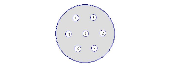
Well # Test A Test B Test C 1 anti-BSA antibody anti-human serum anti-human serum 2 undilute BSA human serum human serum 3 1:2 BSA human albumin monkey serum 4 1:4 BSA human IgG rat serum 5 1:8 BSA goat serum 6 1:16 BSA pig serum 3. Place 40-50 ml of the reagents as indicated in the table into the appropriate wells. Do NOT overfill the wells. For Test A, make dilutions in eppendorf tubes using PBS prior to adding them to the wells.
4. Cover the plate (do not invert), and seal it around the edges with parafilm. Incubate 48 hours at room temperature.
5. On Wednesday, examine the plates for formation of precipitin lines. You should look for: a) the effect of dilution of placement of the equivalence zone, b) the relationship of human serum components, c) the relationship of serum proteins between species.
Study Questions
1. In what situation would you expect to see more than one precipitiatin line between wells on an Ouchterlony plate?
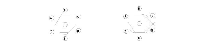
2. The labels from four bottles (A, B, C, and D) of hapten-carrier conjugates were accidentally removed. However, it was known that each bottle contained either hapten 1- carrier 1 (H1-C1), hapten 1 carrier 2 (H1-C2), hapten 2 carrier 1 (H2-C1), or hapten 2 carrier 2 (H2-C2). Ouchterlony assays with either anti-H1-C2 or anti H2-C2 were performed. From the precipitin patterns shown below, determine which conjugate is in each bottle.
Anti-H1-C2 in central well Anti-H2-C2 in central well
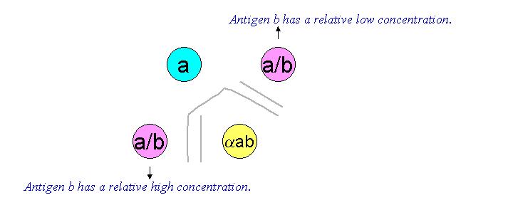
3. You did an Ouchterlony test and got the following result. Which antigen has a higher concentration?